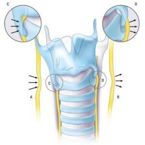
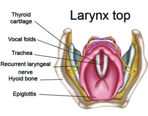
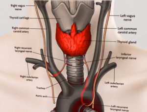
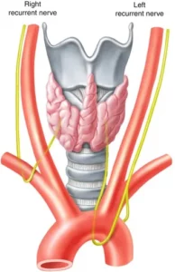
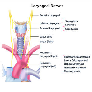
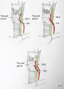
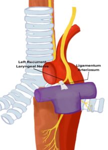
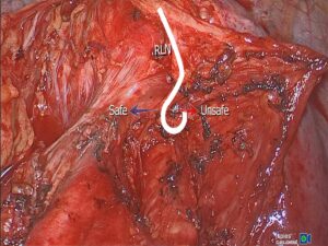
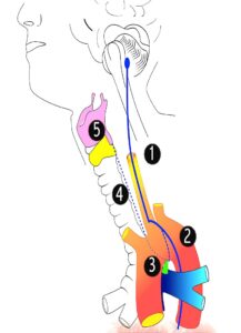
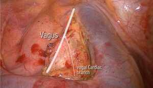
解剖 喉返神经 sunxy md 2025年11月3日 2025年11月3日 RLN(Recurrent Laryngeal Nerve)是非常重要的解剖结构,必须精准辨认 The image illustrates various forms of nerve compression and stretching in the neck. It shows compression of the right vagus nerve by soft tissue (A) and crushing of the recurrent laryngeal nerve by cartilage (C). The left vagus nerve is stretched due to neck hyper-extension (B), while the left recurrent laryngeal nerve is tethered (D). The image also highlights the tilting of the thyroid cartilage and provides enlarged views of the crico-thyroid articulation, indicating the areas where the nerves are most vulnerable to damage.Access to the right recurrent laryngeal nerve from posterior approach and its relation to the right inferior thryoid artery and nearby lymph nodes. Surgical point of view from a robotics surgery procedure.